Subcutaan vetweefsel FDG-opname correleert met systemische ontsteking bij borstkanker
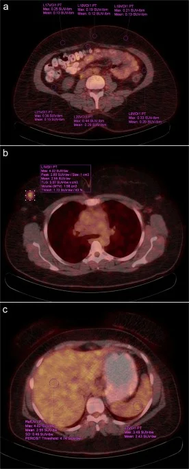
Een retrospectieve single-center cohortstudie (n=358) onderzocht of de FDG-opname in subcutaan vetweefsel op PET/CT correleert met systemische ontstekingsindices, onafhankelijk van de metabole tumorlast bij patiënten met borstkanker.
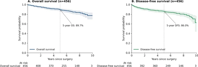
Hogere vetweefsel-opname bleef onafhankelijk geassocieerd met een verhoogde neutrofiel-lymfocytenratio (β=0,0798; p=0,019), met een stijging van ongeveer 9 procent per standaarddeviatie. Deze associatie was niet afhankelijk van metastatische status en niet significant voor de overleving (aHR=1,086; p=0,481).
De bevindingen suggereren dat de opname een weefselspecifieke afspiegeling is van het systemische ontstekingsfenotype, maar vormt geen zelfstandige klinische biomarker. Externe validatie is nodig voordat toepassing in de dagpraktijk overwogen kan worden.
Abstract (original)
PURPOSE: Adipose tissue may contribute to systemic inflammation in breast cancer. This study evaluated whether visceral adipose tissue (VAT) and subcutaneous adipose tissue (SAT) [18F]fluorodeoxyglucose ([18F]FDG) uptakes on positron emission tomography/computed tomography (PET/CT) are associated with inflammatory indices after adjustment for PET-derived tumor metabolic burden and whether metastatic status modifies these associations. MATERIALS AND METHODS: In this retrospective single-center cohort (n = 358) undergoing baseline [18F]FDG PET/CT, metastatic status was defined using an internal three-level clinical stage grouping: early-stage or locally advanced disease was classified as M0 and metastatic disease was classified as MET. VAT and SAT FDG uptakes were quantified as z-scored mean standardized uptake values normalized to lean body mass (SULmean) using the James lean body mass equation. Inflammatory indices from complete blood counts were log-transformed. Prespecified linear regression models with heteroskedasticity-consistent type 3 (HC3) robust standard errors were adjusted for age, body mass index (BMI), fasting glucose, stage, molecular subtype, and PET-derived primary-tumor metabolic burden metrics. Effect modification was tested via exposure × MET interaction. Sensitivity analyses used total-body tumor burden metrics in MET and recalculated SUL using the Janmahasatian lean body mass equation; overall survival (OS) was explored with Cox models. RESULTS: Of 358 patients, 284 (79.3%) were M0 and 74 (20.7%) were MET. In M0 (complete-case n = 267), higher SAT uptake was independently associated with higher log-transformed neutrophil-to-lymphocyte ratio (log(NLR)) (β = 0.0798; 95% confidence interval (CI) 0.0130-0.1466; p = 0.019). In pooled interaction models (n = 323), SAT remained associated with log(NLR) (β = 0.0892; 95% CI 0.0196-0.1588; p = 0.012; ≈9% higher NLR per 1-standard deviation (SD)), with no evidence of effect modification by metastatic status (SAT × MET interaction p = 0.594). Results were consistent after total-body tumor-burden adjustment in MET and after recalculation of SUL using the Janmahasatian lean body mass equation. SAT was not significantly associated with OS (adjusted hazard ratio (HR) = 1.086; 95% CI 0.864-1.364; p = 0.481; n = 346). CONCLUSIONS: Higher SAT FDG uptake was modestly associated with higher NLR independent of tumor metabolic burden, with no evidence of heterogeneity by metastatic status. These findings suggest a potential tissue-specific imaging correlate of host inflammatory phenotype rather than a stand-alone clinical biomarker. Sensitivity and survival analyses remain hypothesis-generating and require external validation.
Dit artikel is een samenvatting van een publicatie in Molecular imaging and biology. Voor het volledige artikel, alle details en referenties verwijzen wij u naar de oorspronkelijke bron.
Lees het volledige artikelDOI: 10.1007/s11307-026-02138-0