T1-mapping extracellulaire volumefractie onderscheidt stroma-rijke rectumtumoren
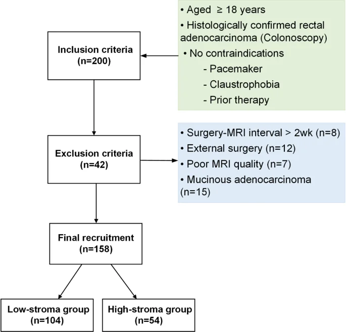
Een prospectieve studie van 158 patiënten met rectumadenocarcinoom onderzocht de waarde van extracellulaire volumefractie (ECV) afgeleid van T1-mapping voor het niet-invasief bepalen van de tumor-stroma verhouding.
ECV vertoonde een matige correlatie met het histopathologische stroma-percentage (ρ = 0,520, p < 0,001) en onderscheidde stroma-rijke tumoren goed (AUC 0,819). Diffusie-gewichtte beeldvorming (ADC) voegde geen significante diagnostische waarde toe.
Deze bevindingen suggereren dat T1-mapping-gebaseerde ECV een reproduceerbare niet-invasieve biomarker kan zijn voor het stratificeren van rectumtumoren, wat relevant is voor prognose-inschatting en behandelkeuzes in de neoadjuvante setting.
Abstract (original)
PURPOSE: To investigate the value of extracellular volume fraction (ECV) derived from T1 mapping and apparent diffusion coefficient (ADC) for non-invasive assessment of tumor stroma percentage (TSP) in rectal cancer. METHODS: This prospective study enrolled 158 patients (104 men, 54 women; mean age 65.96 ± 10.87 years) with rectal adenocarcinoma. All patients underwent 3.0T MRI including pre- and post-contrast T1 mapping and diffusion-weighted imaging. ECV was calculated from native and post-contrast T1 values. TSP was histopathologically assessed on surgical specimens. Interobserver reproducibility, correlations with TSP, diagnostic performance for stroma-rich tumors (TSP > 50%), and associations with clinicopathological features were evaluated. RESULTS: The pre- and post-contrast T1 measurements used to calculate ECV showed excellent interobserver reproducibility (ICC = 0.978 and 0.964, respectively). ECV showed a moderate positive correlation with TSP (ρ = 0.520, P < 0.001), whereas ADC showed no significant correlation (ρ = 0.070, P > 0.05). ECV demonstrated good discrimination of stroma-rich tumors (AUC, 0.819; 95% CI 0.742-0.897), whereas ADC derived from the two-b-value DWI protocol did not discriminate between stroma-rich and stroma-poor tumors (AUC, 0.495; 95% CI: 0.398-0.592; P < 0.001 vs. ECV). Adding ADC to ECV did not significantly improve diagnostic performance compared with ECV alone (AUC = 0.845, P > 0.05; DeLong P > 0.05). ECV differed across tumor differentiation grades after correction (P < 0.05), but not according to LVI status (P > 0.05). CONCLUSION: T1 mapping-derived ECV was reproducible and showed a moderate association with histological TSP, with good performance for distinguishing stroma-rich from stroma-poor tumors.
Dit artikel is een samenvatting van een publicatie in Abdominal radiology (New York). Voor het volledige artikel, alle details en referenties verwijzen wij u naar de oorspronkelijke bron.
Lees het volledige artikelDOI: 10.1007/s00261-026-05769-7